Senior Consultant & Interventional Cardiologist
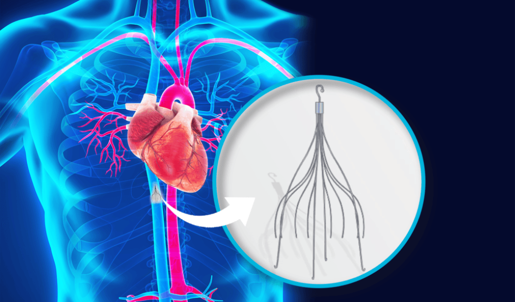
IVC (Inferior Vena Cava) Filter Placement is a minimally invasive interventional procedure performed to help prevent life-threatening pulmonary embolism in patients who are at high risk of blood clots traveling from the legs to the lungs. The inferior vena cava is the body’s largest vein, carrying blood from the lower half of the body back to the heart. An IVC filter is a small, cage-like medical device that is carefully placed inside this vein to capture large blood clots before they reach the lungs.
This procedure is commonly recommended for patients who cannot safely take blood-thinning medications, have experienced recurrent blood clots despite anticoagulant therapy, or require temporary protection during high-risk situations such as major surgery or trauma. Performed under image guidance by an experienced interventional radiologist, IVC filter placement offers a safe, effective, and minimally invasive solution with faster recovery and minimal discomfort.
IVC filter placement is generally considered for patients who are at increased risk of developing deep vein thrombosis (DVT) or pulmonary embolism (PE). Blood clots can form due to prolonged immobility, major surgeries, traumatic injuries, inherited clotting disorders, certain cancers, pregnancy, obesity, or advanced age. Individuals who have had previous episodes of DVT or pulmonary embolism are also at higher risk of recurrence.
In some situations, blood-thinning medications may not be suitable because of active bleeding, recent surgery, severe head injuries, or other medical conditions that increase the risk of hemorrhage. In these cases, an IVC filter provides an additional layer of protection by reducing the chance of dangerous clots reaching the lungs.
Patients requiring IVC filter placement may present with symptoms related to deep vein thrombosis or pulmonary embolism. Deep vein thrombosis commonly causes swelling in one leg, pain or tenderness, warmth, redness, or skin discoloration. Pulmonary embolism may lead to sudden shortness of breath, chest pain that worsens with breathing, rapid heartbeat, dizziness, coughing, or coughing up blood.
It is important to note that some blood clots may not produce noticeable symptoms until complications develop. Individuals with known clotting disorders or significant risk factors should seek timely medical evaluation even if symptoms appear mild.
A comprehensive clinical evaluation is essential before recommending IVC filter placement. The diagnosis begins with a detailed medical history, assessment of clotting risk factors, and physical examination. Imaging studies such as Doppler ultrasound are commonly used to detect deep vein thrombosis, while CT pulmonary angiography helps identify pulmonary embolism. Additional investigations, including blood tests and clotting profile assessments, may also be performed to determine the underlying cause and overall health status.
Advanced imaging guidance, including fluoroscopy or ultrasound, is used during the procedure to accurately position the filter within the inferior vena cava, ensuring optimal effectiveness and safety.
The primary treatment for blood clots remains anticoagulant medication whenever it is safe and appropriate. However, IVC filter placement becomes an important treatment option when blood thinners are contraindicated, ineffective, or temporarily unsuitable. The procedure is typically performed through a small puncture in the neck or groin using a thin catheter under local anesthesia and imaging guidance.
Depending on the patient’s condition, either a permanent or retrievable IVC filter may be selected. Retrievable filters are often removed once the risk of pulmonary embolism decreases or when anticoagulant therapy can be safely resumed. In many cases, IVC filter placement is combined with other treatments, including anticoagulation, catheter-directed thrombolysis, or thrombectomy, to achieve the best possible outcome.
Recovery following IVC filter placement is generally quick, with most patients returning home on the same day or after a short period of observation. Patients are advised to keep the insertion site clean, avoid strenuous physical activity for a brief period, and follow all medication instructions provided by their healthcare team.
Regular follow-up appointments are important to monitor the position and function of the filter through appropriate imaging studies. For patients with retrievable filters, timely evaluation is essential to determine the appropriate time for removal, minimizing the risk of long-term complications while ensuring continued protection against pulmonary embolism.
IVC filter placement is considered a safe and well-established procedure when performed by experienced specialists. However, like any medical intervention, it carries certain risks. These may include minor bleeding or bruising at the catheter insertion site, infection, allergic reactions to contrast material, filter migration, filter fracture, blockage of the inferior vena cava, or difficulty retrieving temporary filters if left in place for an extended period.
Although serious complications are uncommon, regular follow-up imaging and adherence to medical advice significantly reduce potential risks and help ensure the long-term success of the treatment.
Patients should seek immediate medical attention if they experience sudden chest pain, difficulty breathing, coughing up blood, severe swelling or pain in the legs, persistent bleeding from the procedure site, fever, or signs of infection following IVC filter placement. Individuals with a history of deep vein thrombosis, pulmonary embolism, or conditions that increase the risk of blood clots should consult a specialist promptly for evaluation and personalized treatment recommendations.
Early diagnosis, appropriate intervention, and ongoing follow-up play a crucial role in preventing serious complications associated with venous blood clots. Under the expert care of Dr. Sagar Chandrashekhar Bhuyar, patients receive comprehensive evaluation, advanced image-guided treatment, and individualized care to ensure optimal outcomes and long-term vascular health.
Patients choose Dr. Sagar Chandrashekhar Bhuyar as their Consultant Interventional Cardiologist in Secunderabad for his combination of over 20 years of hands-on interventional experience, board certification (MBBS, MD, DNB – Cardiology), international fellowships (FACC, FESC), and an academic role that keeps his practice aligned with current cardiology research.
From preventive cardiology and cardiac imaging to complex coronary interventions, Dr. Bhuyar provides comprehensive heart care tailored to each patient's needs. His expertise includes Angiography, Angioplasty (PCI), Pacemaker Implantation, Peripheral Interventions, Balloon Valvoplasty, and Echocardiography.
Copyright © 2026 Dr. Sagar Chandrashekhar Bhuyar. All Rights Reserved.
WhatsApp us