Senior Consultant & Interventional Cardiologist
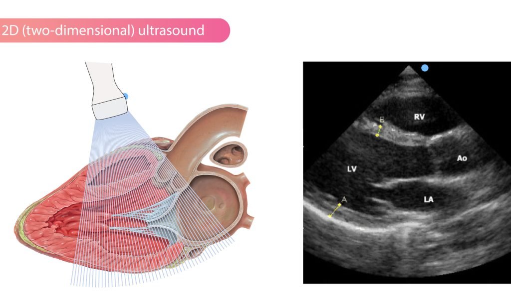
A 2D Echo (Two-Dimensional Echocardiography) is a safe, painless, and non-invasive ultrasound test that provides detailed images of the heart’s structure and function. Using high-frequency sound waves, this advanced diagnostic procedure allows your cardiologist to assess the size of the heart chambers, the movement of the heart muscles, the condition of the heart valves, and the efficiency of blood flow through the heart. Unlike tests that involve radiation, a 2D Echo is completely radiation-free and can be safely performed on patients of all ages.
At Dr. Sagar Chandrashekhar Bhuyar’s practice, 2D Echo plays a vital role in the early diagnosis and ongoing monitoring of various heart conditions. The test helps detect abnormalities before symptoms become severe, enabling timely treatment and better long-term heart health.
A 2D Echo is recommended when there is a need to evaluate the heart’s structure or investigate symptoms that may indicate an underlying cardiac condition. It is commonly advised for patients experiencing chest pain, breathlessness, palpitations, dizziness, fainting episodes, or unexplained fatigue. Individuals with high blood pressure, diabetes, congenital heart disease, previous heart attacks, heart murmurs, or a family history of heart disease may also require a 2D Echo as part of their comprehensive cardiac assessment.
Additionally, this test is frequently used to monitor patients with known heart valve disorders, heart failure, cardiomyopathy, infections affecting the heart, or those recovering after cardiac procedures or surgeries.
A 2D Echo may be recommended if you experience persistent chest discomfort, shortness of breath during daily activities or at rest, rapid or irregular heartbeat, swelling in the legs or ankles, unexplained fatigue, dizziness, fainting spells, or reduced exercise tolerance. These symptoms may indicate problems with the heart’s pumping ability, valve function, or overall cardiovascular health. Even in the absence of symptoms, individuals with significant cardiovascular risk factors may benefit from this diagnostic test for early detection of heart disease.
During a 2D Echo, a trained cardiac technician applies a special gel to the chest and gently moves a handheld ultrasound probe over the skin. The probe emits sound waves that create real-time images of the heart on a monitor, allowing the cardiologist to evaluate the heart’s anatomy and performance. The procedure typically takes between 20 and 45 minutes, is completely painless, and does not require injections, radiation, or significant preparation in most cases.
The images obtained help diagnose a wide range of conditions, including heart valve diseases, congenital heart defects, weakened heart muscles, enlarged heart chambers, reduced pumping function, fluid accumulation around the heart, blood clots, and abnormalities in heart movement.
The findings from a 2D Echo guide the most appropriate treatment plan based on the specific heart condition diagnosed. Treatment may involve lifestyle modifications, medications to control blood pressure or heart rhythm, cholesterol management, treatment for heart failure, or blood-thinning medications when necessary. In certain cases, patients may require minimally invasive procedures such as angioplasty, valve repair or replacement, or other advanced cardiac interventions.
By providing accurate and detailed information about heart function, a 2D Echo enables your cardiologist to recommend the most effective treatment while monitoring your response over time.
Following treatment, regular follow-up appointments and repeat 2D Echo examinations may be recommended to monitor heart function and evaluate the effectiveness of therapy. Patients are encouraged to follow prescribed medications consistently, maintain a heart-healthy diet, exercise as advised by their cardiologist, manage blood pressure, blood sugar, and cholesterol levels, avoid smoking, and attend routine cardiac check-ups. These measures contribute significantly to improved heart health and reduce the risk of future cardiovascular complications.
A standard transthoracic 2D Echo is considered one of the safest cardiac diagnostic tests available. It is non-invasive, does not involve radiation exposure, and has virtually no known risks or complications. Most patients experience no discomfort apart from mild pressure from the ultrasound probe during the examination. Because of its excellent safety profile, the test can be repeated whenever necessary to monitor heart conditions without causing harm.
You should consult a cardiologist if you experience persistent chest pain, unexplained shortness of breath, irregular heartbeat, dizziness, fainting episodes, swelling of the legs, unusual fatigue, or decreased physical endurance. Individuals with hypertension, diabetes, previous heart disease, congenital heart defects, or a strong family history of cardiovascular disease should also undergo regular cardiac evaluations when advised. Early diagnosis through a 2D Echo can help detect heart conditions at an early stage, allowing prompt treatment and significantly improving long-term heart health and overall quality of life.
Dr. Sagar Chandrashekhar Bhuyar commitment to clinical excellence, innovation, and compassionate treatment helps patients achieve better heart health and improved quality of life.
From preventive cardiology and cardiac imaging to complex coronary interventions, Dr. Bhuyar provides comprehensive heart care tailored to each patient's needs. His expertise includes Angiography, Angioplasty (PCI), Pacemaker Implantation, Peripheral Interventions, Balloon Valvoplasty, and Echocardiography.
Copyright © 2026 Dr. Sagar Chandrashekhar Bhuyar. All Rights Reserved.
WhatsApp us